 About Cephalometrics A to
Z About Cephalometrics A to
Z |
 |
|
How to import color photographs and radiographs Creation of informed consent document |
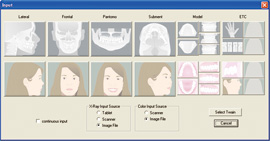
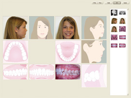
How to import color photographs and radiographs |
 |
||
 |
 |
|
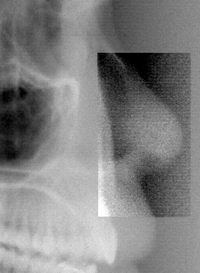
Images or X-rays can be enhanced and made clearer by using the "enhance" function. The scanned image to be enhanced is then boxed, and the enhance function selected.
Image too dark, too bright, not sharp enough? Correct it immediately
on your screen! Image Rotation Copy and Paste Image Integration |
 |
|
Explaining treatment plans to the patient is made easier with the model analysis function. It can calculate the space discrepancy using the Bezier curve. It can also simulate tooth extractions. |
||
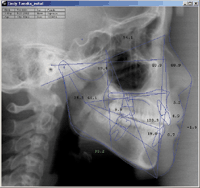
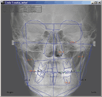
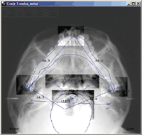
Lateral analyses include Ricketts, McNamara, Steiner(Tweed), Jarabak, Roth, Sassouni, Downs, Holdaway,Burstone,and four user defined combinations and variations analysis. Frontal analysis: Ricketts, symmetry and four user defined analysis. Submentovertex analysis: Submentovertex and four user defined analysis. |
||
 �@ �@ �@ �@ |
||
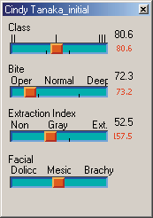
Your treatment (relapse, ODI and CF, open bite tendency andextraction versus non-extraction) is standardized or judged against the APDI of Dr. Kim, who has a clinical experience of over 4,000 cases. This summaries the diagnosis into text form, and diagnosis window. |
 |
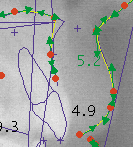
The soft-tissue and hard-tissue outlines are corrected with a Bezier curve which can be finely adjusted. |
 |
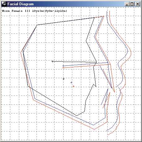
You can display common analyses such as superposition by the standard value, superposition by the SN-FH plane, and display of soft-tissue. Dowens-North Western analysis and linear analysis can also be displayed in this manner. These can also be represented in polygonal form. |
 |
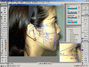
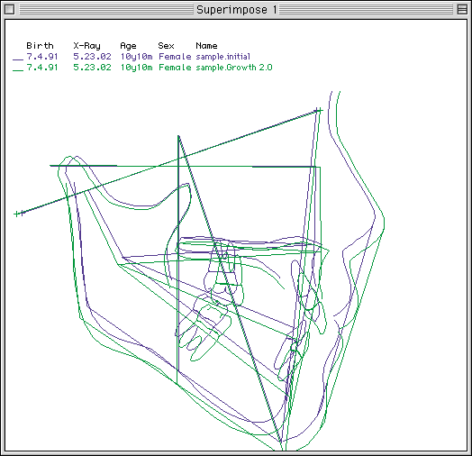
Easily superimpose cephalometric tracing over the patient's photo. Superimpose tracings of different timepoints with standard superimposition references (SN at Sella, Frankfort at Porion, Na-Pg at ANS-PNS....). |
 |